SM Increases the Potential Difference of Renal Tubule
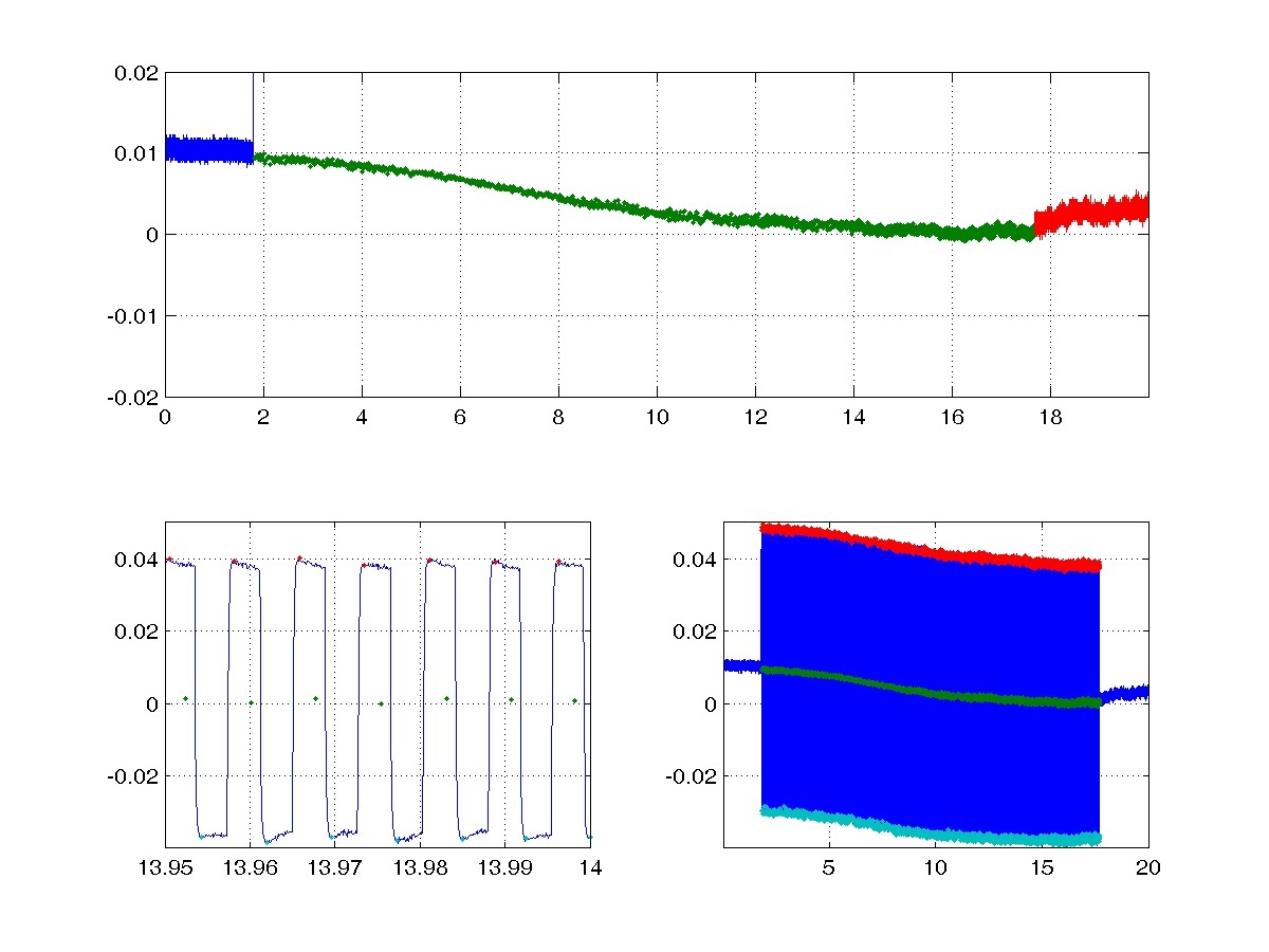
We conducted in vivo study of the synchronization modulation electric field on functions of living kidney of rats by monitor the field-induced changes in the transepithelial potential difference (TEPD) on proximal convoluted tubules. Before the field application, the rest TEPD is about 11 mV shown in the lower right panel. The lower left panel of the figure below shows the detailed changes for the field-induced, pulsed TEPD measured at about 14th second. Initially, the field-induced membrane potential oscillates symmetrically with respect to the rest TEPD. As the field frequency gradually increases, profile of the oscillating TEPD is shifted to the negative direction until removal of the electric field. The averaged value of the oscillating cycles are calculated and plotted as the middle green curve and redrawn in the upper panel. Clearly, in response to the SM electric field, the TEPD gradually becomes more negative. The absolute value of TEPD measurements may not be accurate because of the electrode junction potential. The 10 mV shift of the TEPD in the negative direction clearly indicates that more cations are transported from lumen to the peritubular space.
Selected Manuscript
- Mathis, C., Fang, ZH. Chen, W., Synchronization modulation electric field can effectively increase the trans-epithelim potential difference in proximal tubule in kidney of rats (submitted).